Download PDF – Human Nervous System: Anatomy, Physiology & Exam Notes

The nervous system is the master controlling and communicating system of the human body. Every thought, action, sensation, and emotion reflects its rapid electrical and chemical signaling activity.
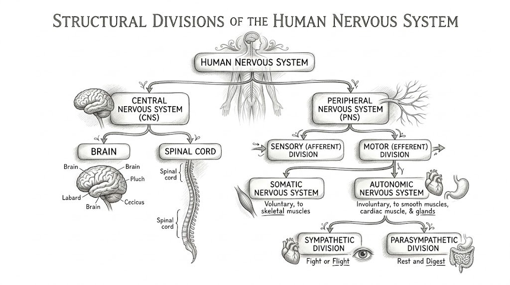
1. Structural and Functional Organization
To understand the nervous system, it is categorized structurally (by location) and functionally (by direction of signaling and target tissue control).
A. Structural Divisions
- Central Nervous System (CNS): Consists of the Brain and the Spinal Cord. It occupies the dorsal body cavity and acts as the integration and control center.
- Peripheral Nervous System (PNS): Consists of nerves emerging from the brain and spinal cord (12 pairs of cranial nerves and 31 pairs of spinal nerves) and ganglia.
B. Functional Subdivisions of the PNS
- Sensory (Afferent) Division: Carries nerve impulses from sensory receptors in skin, organs, and muscles toward the CNS.
- Motor (Efferent) Division: Transmits impulses away from the CNS to effector organs (muscles and glands).
- Somatic Nervous System (SNS): Controls voluntary movements of skeletal muscles.
- Autonomic Nervous System (ANS): Controls involuntary responses of smooth muscles, cardiac muscle, and glands.
- Sympathetic Division: Mediates the “Fight-or-Flight” response during stress.
- Parasympathetic Division: Mediates the “Rest-and-Digest” activities, promoting conservation and maintenance.
Examiner’s Warning/Pitfall: Always remember that sensory/afferent fibers enter via the dorsal horn of the spinal cord, whereas motor/efferent signals exit via the ventral horn (Mnemonic: DAVE — Dorsal Afferent, Ventral Efferent).
2. Histology: Neurons and Neuroglia
Nervous tissue consists of two primary cell populations: excitable signaling cells (neurons) and supportive non-conducting cells (neuroglia).
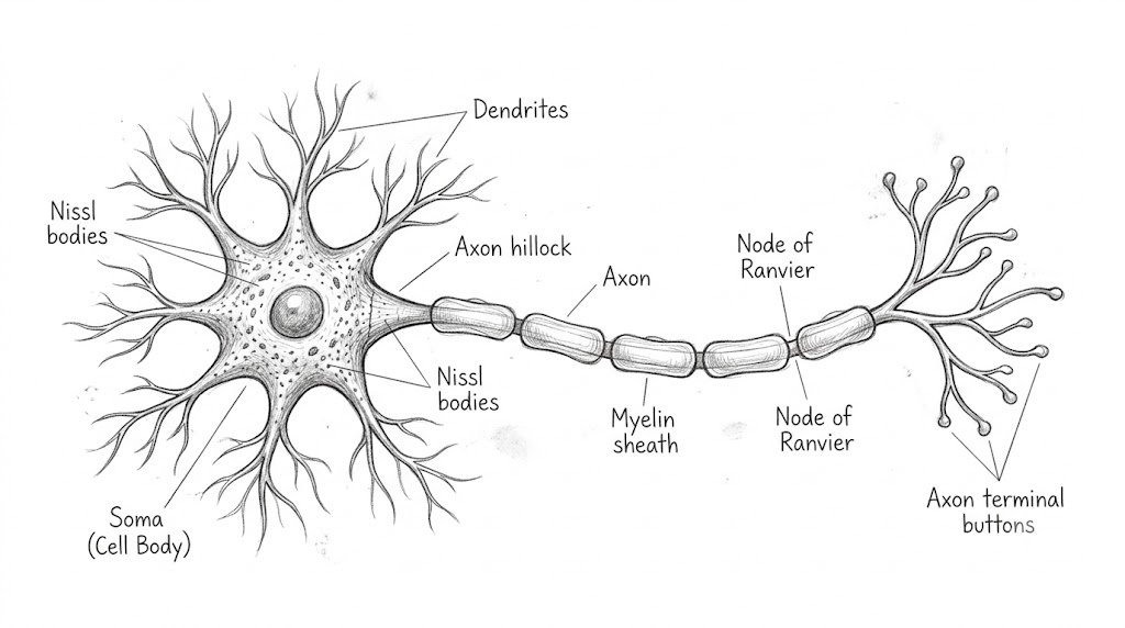
A. The Neuron (Functional Unit)
- Dendrites: Receptive branch-like extensions that receive chemical signals from adjacent neurons.
- Cell Body (Soma/Perikaryon): Contains the nucleus and metabolic organelles, including prominent clusters of rough endoplasmic reticulum known as Nissl bodies (sites of high protein synthesis).
- Axon Hillock: Cone-shaped elevation where the axon originates and action potentials are triggered.
- Axon: Long, slender projection responsible for transmitting electrical impulses away from the soma.
- Myelin Sheath: Insulative lipoprotein wrap that speeds up impulse transmission via saltatory conduction. Formed by Oligodendrocytes in the CNS and Schwann cells in the PNS.
- Nodes of Ranvier: Unmyelinated gaps along the axon rich in voltage-gated sodium channels.
B. Neuroglial Cells (Supportive Cells)
| Glial Cell Type | Location | Key Function | High-Yield Exam Point |
|---|---|---|---|
| Astrocytes | CNS | Form blood-brain barrier (BBB), regulate extracellular potassium and neurotransmitter levels. | Most abundant glial cell in the brain. |
| Oligodendrocytes | CNS | Produce myelin sheaths around multiple axons simultaneously. | Damaged in Multiple Sclerosis. |
| Microglia | CNS | Phagocytize cellular debris, microbes, and damaged tissue. | Macrophage-lineage immune defense of the CNS. |
| Ependymal Cells | CNS | Line brain ventricles and central canal; circulate cerebrospinal fluid (CSF) via cilia. | Part of the choroid plexus synthesizing CSF. |
| Schwann Cells | PNS | Form myelin sheaths around single segments of peripheral axons; assist axon regeneration. | Damaged in Guillain-Barré Syndrome. |
| Satellite Cells | PNS | Surround and cushion neuron cell bodies in peripheral ganglia. | Analogous in function to CNS astrocytes. |
3. Neurophysiology: Resting Membrane Potential & Action Potential
Neurons communicate via transient electrical fluctuations across the plasma membrane driven by selective ionic conductances.
A. Resting Membrane Potential (RMP)
- Typical neuronal RMP is approximately −70 mV (inside negative relative to outside).
- Maintained primarily by high resting permeability to K+ (via leakage channels) and the electrogenic Na+/K+ ATPase pump (pumps 3 Na+ out for every 2 K+ in).
B. Phases of the Action Potential
- Resting State: Voltage-gated Na+ and K+ channels remain closed. Membrane potential sits at −70 mV.
- Depolarization: A stimulus reaches threshold (approx. −55 mV), prompting rapid opening of activation gates of voltage-gated Na+ channels. Rapid Na+ influx drives membrane potential up to +30 mV.
- Repolarization: Inactivation gates of Na+ channels close; voltage-gated K+ channels open. Potassium rushes out down its electrochemical gradient, returning potential toward negative values.
- Hyperpolarization (After-potential): Prolonged efflux of K+ because potassium channels close slowly, dropping the potential slightly below resting level (−80 to −90 mV) before settling back via the Na+/K+ pump.
High-Yield Exam Focus:Absolute Refractory Period occurs during depolarization and early repolarization when Na+ channels are inactivated; NO stimulus can produce a new action potential. Relative Refractory Period follows, during which an exceptionally strong suprathreshold stimulus can generate an impulse.
4. Synaptic Transmission
A synapse is the functional junction transmitting signals between an axon terminal and an effector cell or adjacent dendrite.
- Action potential reaches the presynaptic axon terminal.
- Membrane depolarization triggers voltage-gated Ca2+ channels to open.
- Ca2+ influx triggers synaptotagmin/SNARE complex-mediated exocytosis of neurotransmitters into the synaptic cleft.
- Neurotransmitter binds to postsynaptic receptors, inducing graded potentials:
- EPSP (Excitatory Postsynaptic Potential): Typically involves Na+ influx (e.g., Acetylcholine, Glutamate).
- IPSP (Inhibitory Postsynaptic Potential): Involves Cl− influx or K+ efflux (e.g., GABA, Glycine).
5. Comparative Anatomy: Sympathetic vs. Parasympathetic Nervous System
| Feature | Sympathetic Division | Parasympathetic Division |
|---|---|---|
| Origin / Outflow | Thoracolumbar (T1 – L2 segments) | Craniosacral (Cranial nerves III, VII, IX, X and S2 – S4) |
| Ganglia Location | Close to spinal cord (Paravertebral / Prevertebral chain) | Near or inside target organs (Terminal intramural ganglia) |
| Preganglionic Fiber | Short (releases Acetylcholine) | Long (releases Acetylcholine) |
| Postganglionic Fiber | Long (releases Norepinephrine, except sweat glands: ACh) | Short (releases Acetylcholine) |
| Pupil Response | Mydriasis (dilation) | Miosis (constriction) |
| Heart Rate & Bronchi | Increases heart rate; bronchodilation | Decreases heart rate; bronchoconstriction |
| GI Motility & Secretions | Inhibited / decreased | Stimulated / increased |
Quick Memory Mnemonic: Parasympathetic cranial nerves = 1973 (Cranial nerves X [10], IX [9], VII [7], III [3]).
6. Summary / Key Takeaways for Competitive Exams
- Glutamate is the primary excitatory neurotransmitter of the CNS; GABA is the primary inhibitory neurotransmitter in the brain.
- Saltatory conduction occurs at the Nodes of Ranvier, drastically saving ATP and increasing conduction velocity.
- The Cranial Nerve X (Vagus nerve) provides approximately 75% of all parasympathetic outflow to thoracic and abdominal viscera.
How to Download Human Nervous System: Anatomy, Physiology & Exam Notes PDF?
- Wait for 1 Minute on this page until the download button unlocks.
- While waiting, we highly recommend exploring our videos or reading the preview above.
- Once the timer ends, the Download Now button will become active.
🎥 Video Resources
Nervous System
Nervous System

The Nervous System, Part 1: Crash Course Anatomy & Physiology #8

Central & Peripheral Nervous System Explained | CNS vs PNS Nursing Guide | Mini Lesson Series

Divisions of the Nervous System - How we divide up the system to better understand it!

Nervous System Basics - Made EASY!

ACE Your Nervous System Exam - Comprehensive Overview!
What You Will Get in Human Nervous System: Anatomy, Physiology & Exam Notes PDF?
- ✔ A high-quality PDF of Human Nervous System: Anatomy, Physiology & Exam Notes with proper formatting
- ✔ Easy to read offline on mobile, tablet, or desktop
- ✔ Great for revision, practice, and quick reference
- ✔ Additional learning support with related videos and resources linked on this page
- ✔ Supports our mission: “Knowledge for Everyone”
Why Choose Solotutes?
At Solotutes, we provide access to notes, quizzes, MCQs, one-liners, and tests for multiple exams. By downloading Human Nervous System: Anatomy, Physiology & Exam Notes in PDF format, you not only get valuable material but also become part of a growing community of learners. We regularly update our posts with videos, explanations, and extra content to help you understand topics better.
FAQs – Human Nervous System: Anatomy, Physiology & Exam Notes Download
Q. Is this PDF really free?
✅ Yes! Free resources are completely free. Premium resources require a one-time unlock or pass.
Q. Do I need to sign up?
❌ For free PDFs, no account is needed. For premium tests, login is required to verify your purchase.
Q. Can I share this PDF with my friends?
✔ Yes, please share and help others learn too.
About Solotutes
Solotutes.com is dedicated to students preparing for competitive exams, schools, and universities. We provide free PDFs, quizzes, MCQs, notes, and one-liners to make learning easy and accessible.
👑 Tired of Waiting and Ads? Go PRO!
Upgrade to the SoloTutes Pro Pass and supercharge your exam preparation.
- 🚫 Ad-Free Experience: Say goodbye to ads and 1-minute waiting timers.
- 📝 Custom PDF Generator: Build your own exam/subject-wise PDFs.
- 📚 PDF Library: Access 1000+ question eBooks.
- 🔓 Unlock Test Series: Get access to all current and upcoming paid test series.